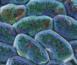


Generation change in the gut
Institute of Anatomy II, Jena University Hospital
At the Institute of Anatomy II, a special imaging technique is used that allows researchers like Prof. Andreas Gebert to make three-dimensional and time-resolved observation of cells: intravital two-photon microscopy. In this photograph, a series of images taken in succession makes the generation change of epithelial cells of intestinal villi visible. Epithelial cells form the boundary between tissue and the cavity of the intestine. Overaged epithelial cells, whose nuclei are shown here in yellow and red, detach from their cell-cluster and pass into the lumen of the small intestine. This makes room for younger cells, which appear in green and blue color. Using such methods, scientists can better understand aging processes in the intestine.
© Andreas Gebert