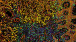


One Step Ahead
Section Pathology, Institute of Forensic Medicine, Jena University Hospital
With this image, apl. Prof. Alexander Berndt gains deeper insights into a malignant tumor growing in a patient’s oral cavity. To create it, he prepared a tissue section in such a way that fluorescent dyes mark different types of cells. Simultaneous imaging of several such dyes is called "multiplex-imaging". Thus, in addition to healthy cells (brown), also the destructively growing tumor cells (yellow), blood vessels (red), and immune cells (green), which are attacking the tumor, can be seen here. Examining such images allows researchers to draw conclusions about the role of different cell types during tumor development. This will allow to make more precise prognosis for this tumor type in the future and to assess the therapy response more accurately.
© Section Pathology / UKJ